
Receiving a diagnosis of hydrocephalus for your child can feel overwhelming.
As Harry’s parents, we had never even heard the word hydrocephalus, before the doctor told us what was going on with Harry.
At the time, we went down quite a scary ‘Google rabbit-hole’, and we want to try and prevent other parents from doing the same.
With that in mind, we’ve put together a written guide to some of the frequently asked questions about the condition.
Please note, we’re not medical professionals, but our pages are put together with support and input from paediatric neurosurgical teams.
This page has information about childhood hydrocephalus, its symptoms, causes, and treatments. We hope this page will be useful for parents and carers of children with hydrocephalus.
The information on this page may also be useful for health professionals such as GPs or health visitors, and education professionals such as teachers.
Hydrocephalus is an abnormal build-up of cerebrospinal fluid (CSF) in the brain.
The excess fluid causes pressure inside the skull, which, in babies under 12 months old, often leads to very fast head growth, or an unusually large head.
In very serious cases, hydrocephalus can cause permanent brain damage, or even be fatal, if it is left untreated.
Although some descriptions can feel frightening, it’s important to remember that many children with hydrocephalus go on to thrive with timely treatment and the right support.
Sometimes the condition is present when a baby is born – this is called congenital hydrocephalus.
Sometimes the condition develops after a baby has been born – this is called acquired hydrocephalus.

Everybody has CSF around their brain and spinal cord. It is a colourless liquid which has several jobs:
Our bodies are always producing new CSF, while the old fluid is absorbed into our blood stream. If something happens to disrupt that process, it can quickly lead to an abnormal build-up of CSF within the brain, called hydrocephalus.
This can happen because:
“Water on the brain” is not an accurate term because we know that CSF is the liquid that causes hydrocephalus, not water. It is quite an outdated way to refer to the condition, but you will find that the term is often still used to describe hydrocephalus in very basic language.
This is because the word hydrocephalus is made up of two Greek words:
Together, the word literally means “water head”.
There are two ways to say hydrocephalus, depending on where you live.
Many children may never know the exact cause of their hydrocephalus, which can be difficult to accept, however, the known causes of hydrocephalus include:
Inside your brain you have spaces which are filled with CSF – these are called ventricles.
Between the ventricles, there are small passageways which allow CSF to flow between them. One of these passageways is called the cerebral aqueduct.
In some children, the aqueduct in their brain is too narrow, which means the flow of CSF slows down or becomes completely blocked.
If CSF cannot escape, it causes the ventricle to swell, leading to hydrocephalus.
This narrowing might not cause symptoms until many years after birth.
This is a condition where the spine does not form properly when the baby is developing in the mother’s womb.
As well as causing problems in the spine, spina bifida can lead to changes in the structure of the brain.
This means most babies born with spina bifida also develop hydrocephalus.
Arachnoid cysts are sacs filled with CSF which can form on your brain or spinal cord. The thin outer layer of the cyst is made from the membrane that covers the brain called the arachnoid membrane.
As fluid builds up inside the arachnoid cyst, it grows like a water balloon – which can sometimes cause a blockage in the flow of CSF and cause hydrocephalus.
Occasionally, genetic changes inherited from parents can lead to hydrocephalus.
For example, a specific mutation in the X chromosome can cause hydrocephalus in boys, known as ‘X‑linked hydrocephalus’.
Another genetic condition called Dandy-Walker syndrome can also cause hydrocephalus.
Bleeds in the brain, known as haemorrhages, can cause blockages in the ventricles, or stop CSF from being re-absorbed, leading to hydrocephalus.
Hydrocephalus can sometimes develop in babies born prematurely (before week 37 of pregnancy). This is thought to be caused by bleeding from the fragile blood vessels inside the brain.
The brain is covered by three thin protective layers. These are membranes, known as the meninges.
Meningitis occurs when those membranes become inflamed because of an infection. It can lead to scarring of the protective layers, which can restrict the flow of CSF, and lead to hydrocephalus.
Tumours can put pressure on the brain and block the flow of CSF, which can cause hydrocephalus. This can even be the case when the tumour is benign, which means non-cancerous, such as a colloid cyst.
To get the most accurate answer to this question, based on your child’s individual circumstances, it’s best to speak to your child’s neurosurgery team.
This is because the outlook for children with hydrocephalus can vary greatly depending on the cause and how soon it was picked up.
Some children with hydrocephalus will have permanent brain damage, while others will have very few problems in the long-term. If left untreated, however, the condition can be fatal.
Although difficult to predict, the potential long-term complications of hydrocephalus can include:
Not all children with hydrocephalus will experience these complications, which is why it’s important to speak with your child’s care team.
While there is no cure for hydrocephalus, it can be managed with treatment. The goal is to drain the build-up of CSF and reduce the pressure on the brain.
There are two main types for treatment for hydrocephalus, which both involve brain surgery:
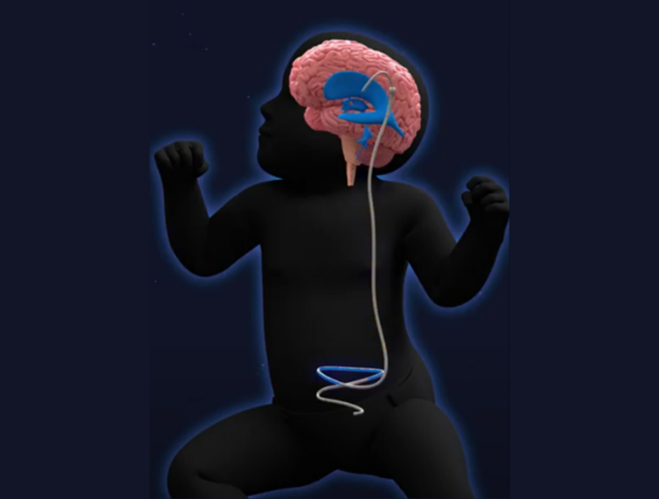
We find the best way to describe a shunt is to compare it to a plumbing system – like the water pipes we have in our homes – simply because it takes fluid from one place to another.
A shunt is a thin, flexible plastic tube which takes the excess fluid away from the brain and transports it to somewhere else in the body to be safely absorbed into the bloodstream.
How will my child’s shunt be fitted?
Shunt surgery is performed in an operating theatre, under a general anaesthetic. The shunt will be implanted inside your child’s body and may take several hours to complete. Hydrocephalus is the most common reason for brain surgery in children, so your neurosurgeon will likely be very experienced in this procedure.
How do shunts work?
One end of the shunt tubing sits in the brain, in the location of the excess fluid. The tubing is connected to a valve which can control the amount of CSF that is drained. This is a one-way valve, which makes sure the CSF flows in the right direction. The remaining tubing on the other side of the valve flows down the neck, (the shape of this is sometimes visible under your child’s skin) and continues to its destination.
There are different types of shunts which drain the fluid into different parts of the body. Your surgeon will advise which type is best for your child:
Most shunt valves also have a reservoir attached, which contains a small amount of CSF. Sometimes, your child’s neurosurgery team may want to perform a ‘shunt tap’ to test if the shunt is working, or to check for signs of infection.
They will use a sterile needle to take some of the CSF from the shunt reservoir, which is typically located under the skin at the back of your child’s head, near to their ear. This test can reveal the pressure of the CSF to show how well the shunt is working, or the CSF can be sent for testing in the lab.
If your child experiences symptoms of a shunt complication or infection, you should contact your child’s healthcare team immediately.
Whilst shunts are a highly effective treatment for hydrocephalus, they can stop working properly over time.
This can happen because of a blockage in the tubing or valve, an infection, fracture or disconnection of the tubing (especially as a child grows), movement of the tubing, and over-drainage of CSF.
Unfortunately, there is no way of knowing how often this will happen for your child. We know of children who have never experienced a shunt malfunction since their first shunt was placed, and we know of children who have experienced dozens of problems with their shunt over the course of their childhood, and into adulthood.
Rough estimates suggest that around 4 in 10 shunts will fail within the first year after surgery, and around 7 out of 10 will need replacing within 10 years.
Remember, every child is different. Your medical team will guide you and monitor your child closely.
If your child’s shunt stops draining properly, the CSF will build-up in the ventricles again, and the pressure around the brain will rise – causing symptoms of hydrocephalus to return.
These symptoms can vary significantly between children and depend on the age of the child, and how quickly the shunt stops working:
Symptoms include, but are not limited to:
You know your child best. If something feels ‘not quite right’, it’s always okay to call your team.
Like any plumbing system, shunts can sometimes block. If scans show that your child’s shunt is blocked, this will be discussed with you and they will likely be sent to the operating theatre as a matter of priority, for a procedure known as a shunt revision, to replace all or part of the shunt.
Shunts may also need to be replaced in an operating theatre if they become infected.
As well as the symptoms of hydrocephalus described above, children with shunt infection will have specific symptoms including, but not limited to:
Infections are most likely to happen within the first 1-3 months of the shunt being implanted. Shunt infections will also require treatment with antibiotics.
An endoscopic third ventriculostomy (known as an ‘ETV’) is an alternative treatment option for some children with hydrocephalus.
It is usually recommended if a child’s hydrocephalus is caused by a blockage within the ventricular system, which prevents the CSF from flowing normally e.g. a tumour, or a cyst.
An ETV is a surgical procedure carried out in an operating theatre, under a general anaesthetic and may take several hours to complete.
Your child’s surgeon will make a small hole in the floor of the third ventricle in their brain. They will do this using a small camera called a neuroendoscope.
This hole creates a new route for the fluid to escape, bypassing the blockage. CSF can then drain out from the third ventricle and into the space surrounding the brain, where it is naturally absorbed into the bloodstream.
An ETV creates a natural alternative pathway for the excess CSF to drain away, without the need for a permanent implant such as a shunt.
In some cases, a successful ETV procedure may eliminate the need for a shunt altogether.
The flow of CSF is usually enough to keep the hole in the third ventricle open. However, if the hole closes over, the ETV may need to be repeated, or – if that is not possible – some children may need to be fitted with a shunt.
Some surgeons also insert a CSF reservoir into the ventricles at the same time as performing an ETV. This reservoir can be used to drain CSF in an emergency (to reduce the pressure), if the ETV stops working.
Your child’s healthcare team will be able to tell you whether ETV is an option for them.
If your child experiences any symptoms of an ETV complication or infection, you should contact your child’s healthcare team immediately.
Complications following an ETV include closure of the hole created during surgery, bleeding, and infection.
If the hole created during surgery closes, much like a shunt that is malfunctioning, the CSF will build-up, the pressure around the brain will rise, and the child will experience hydrocephalus symptoms again.
As before, these symptoms depend on the age of the child, and how quickly the the ETV stops working, but can include:
If your child’s ETV stops draining CSF effectively, they may need surgery to revise it. Some children may also go on to have a shunt fitted.
You know your child best. If something feels ‘not quite right’, it’s always okay to call your team.
You can speak to the healthcare team looking after your child to find out more information about any aspect of hydrocephalus.
More information about hydrocephalus can be found on our signposting page
There are also several hydrocephalus support groups on Facebook for parents and carers. These groups are run by families of children with the condition, and are independent from Harry’s HAT – we are not responsible for the content or information in these groups.